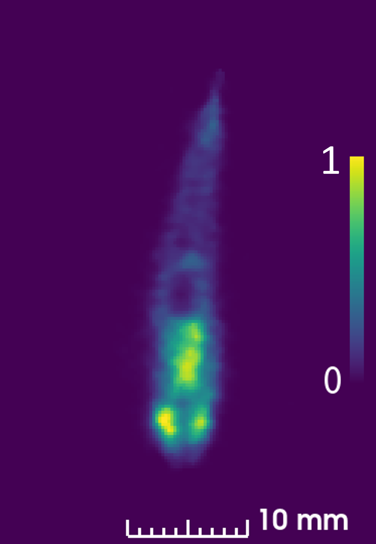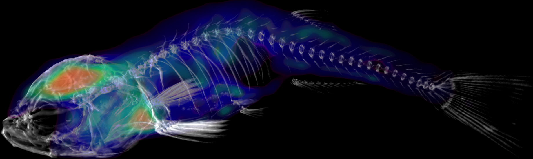
Preclinical positron emission tomography (PET) is an indispensable tool in modern biomedical research. As a highly sensitive, noninvasive molecular imaging technique, it enables the real-time, three-dimensional visualization of biochemical and physiological processes in living organisms.
- Translational relevance: It bridges the gap between in vitro experiments and clinical application in humans, as the same radiotracers and mechanisms can be studied.
- Longitudinal studies (in vivo monitoring): Because the method is noninvasive, the same animals can be studied repeatedly over days, weeks, or months. This drastically reduces the number of animals required (3R principle: Reduction) and increases statistical power, since each animal serves as its own control.
- Highest sensitivity: PET can detect very low concentrations of radiotracers, making functional changes, receptor interactions, or metabolic processes visible even before anatomical changes occur.
Below, you will find a detailed overview of our current research projects. A particular focus of our work is on pioneering research in the field of adult zebrafish PET. Due to their genetic similarity to humans and their excellent suitability for high-throughput screening, adult zebrafish offer a revolutionary, cost-effective, and highly translational model for investigating disease mechanisms and developing therapeutic agents.
For PET imaging, the Isotope Laboratory uses the infrastructure of SAIL as well as that of Prof. Rafecas's Nuclear Imaging working group at the Institute of Medical Engineering (IMT).