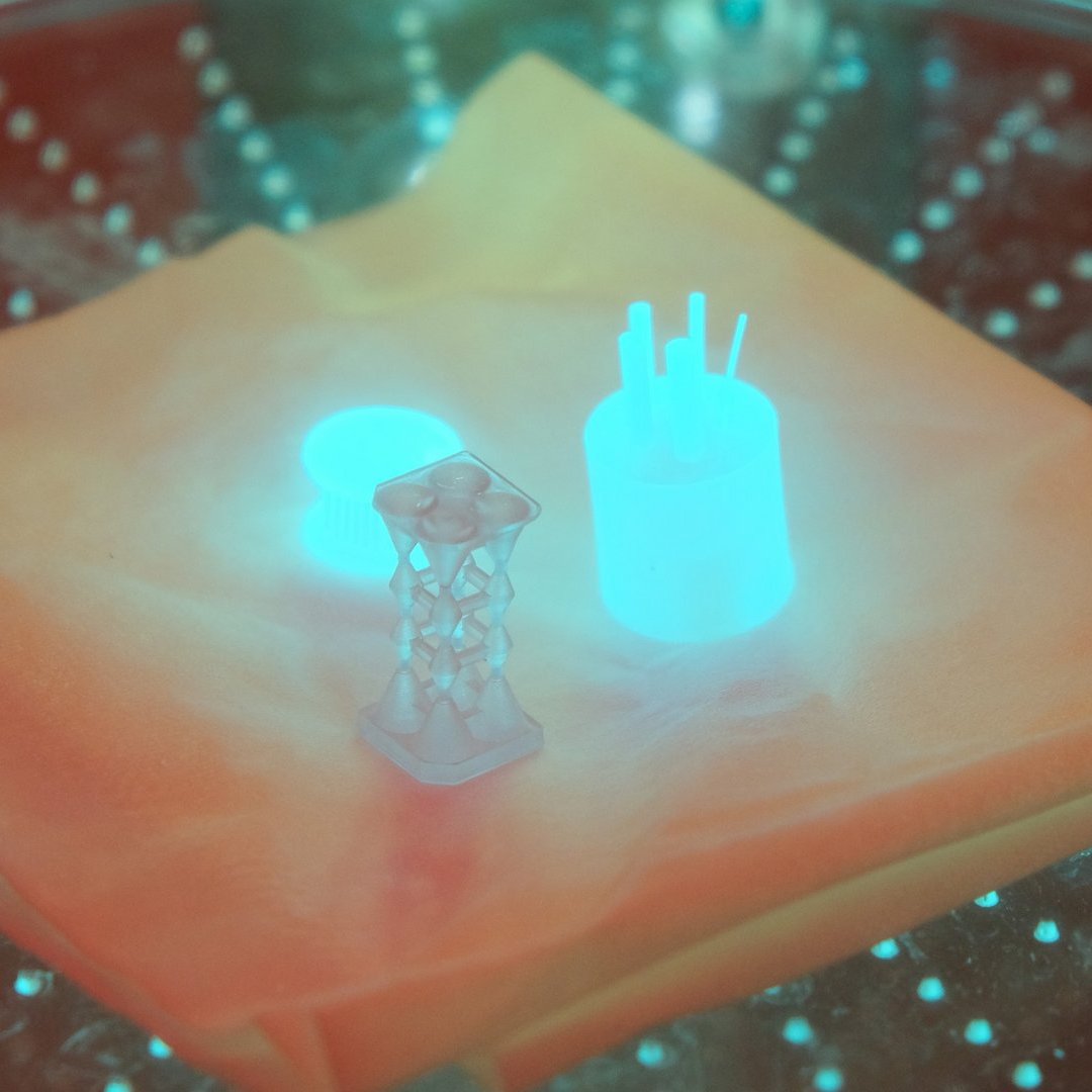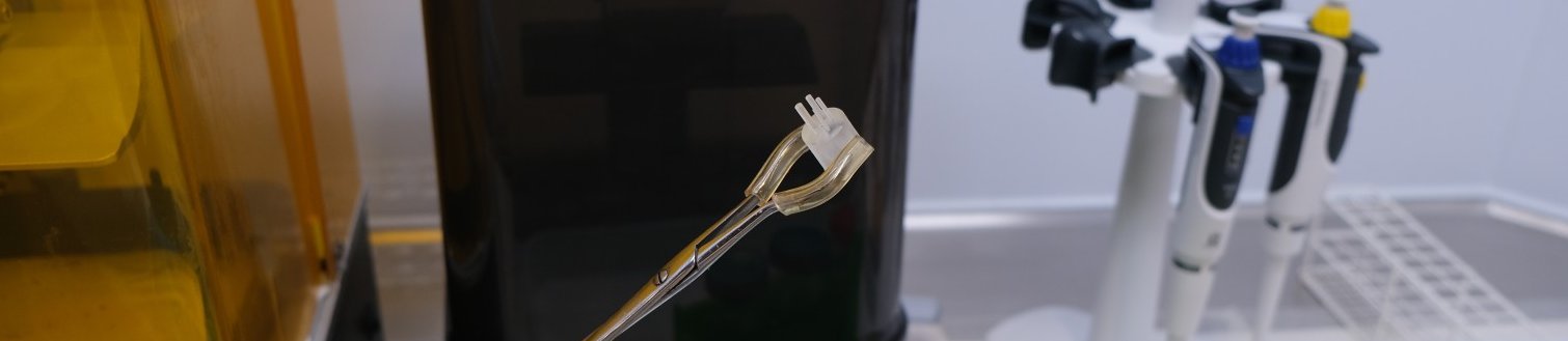
The use of 3D printing for biomedical applications has steadily increased in recent years. Today, complex geometries in virtually any desired shape can be produced in-house using 3D printers for a wide variety of materials, cost-effectively and with high precision. For example, 3D printing enables the production of phantoms that mimic the mechanical properties of the human body, as well as phantoms for quality assurance or evaluation of medical imaging systems such as X-ray, computed tomography (CT), single-photon emission computed tomography (SPECT), and positron emission tomography (PET). In nuclear imaging—that is, in scintigraphy, SPECT, and PET—a wide variety of phantoms are typically used for quality assurance and system performance evaluation. These phantoms are either solid or consist of various empty cavities that can be filled with radiotracers or solutions containing specific radioisotopes. The design may comply with specific standards, such as the National Electrical Manufacturers Association (NEMA) NU 2-2018 and NEMA NU 4-2008 for clinical and preclinical PET, respectively. A common challenge with these liquid-filled phantoms (whether conventional or 3D-printed) is often their leak-tightness and the filling process.
To address these issues, we have collaborated with the Nuclear imaging research group at the Institute of Medical Enineering to develop a method for producing radioactive phantoms that no longer require a filling agent.
We are already able to use a wide range of radionuclides for PET imaging. In the long term, we aim to expand this method to include long-lived nuclides as well as other modalities of nuclear imaging.
Together with Fraunhofer IMTE, we are also currently working toward commercializing these phantoms.